Fluorescent Fireworks 3
Fluorescent Fireworks 3
Abigail Lytton-Jean, Giovanni de Nola, Laurie Boyer, John Day
Koch Institute at MIT
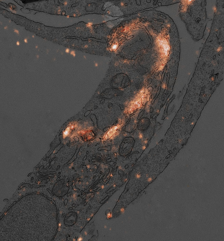
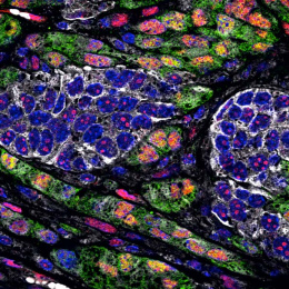
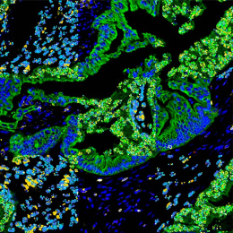
This electron microscopy image shows a section of a cell in which lysosomes were labeled with fluorescent antibodies using a specialized technique developed in our lab. The darker background reveals the exquisitely preserved cellular architecture, while the glowing areas highlight individual lysosomes tethered throughout the cytoplasm.
The contrast between light and shadow was intentionally designed to let the fluorescent signal emerge vividly—like fireworks against a night sky, or the silhouette of a flaming dragon rising from the cell’s interior. The image reflects the vitality of the cell itself: a glimpse of molecular activity captured in form and light.