Engineering the Negative Space
Engineering the Negative Space
Vardhman Kumar, Joa Yun, Heather Fleming, Sangeeta Bhatia
Koch Institute at MIT
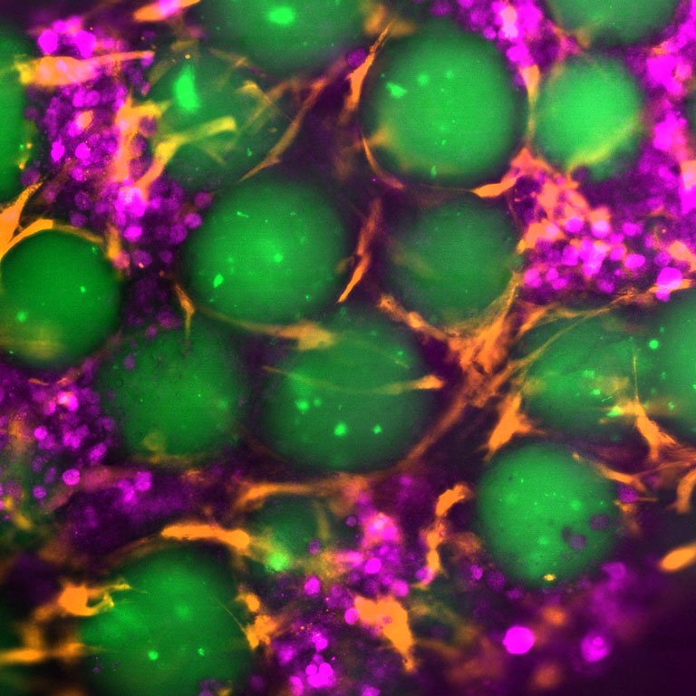
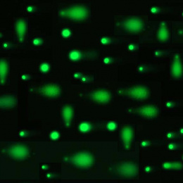
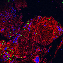
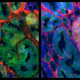
This image illustrates a 3D liver tissue construct consisting of hydrogel microspheres (green), hepatocytes (magenta), and supporting fibroblasts (yellow). The hydrogel provides a matrix that mimics the liver’s extracellular environment, organizing cells into functional architectures and enabling nutrient exchange. Notice the cells in the spaces between the green microspheres, the magenta hepatocytes clustering along the tiny corridors and the yellow fibroblasts lining and remodeling these channels. It’s like a cup of boba; pack the pearls together and the gaps between them become hallways where the cells move, communicate, and self-assemble. By engineering the “negative space”, the interconnected voids formed between the microspheres, the system intentionally sculpts pore size, connectivity, and confinement to guide self-assembly, polarization, and cell migration. Fibroblasts contribute extracellular matrix proteins and paracrine factors that support hepatocyte viability and promote tissue remodeling.