A Zebrafish Model of Uveal Melanoma 3
A Zebrafish Model of Uveal Melanoma 3
Submitted by Dahlia Perez of the Lees Laboratory at the Koch Institute for Integrative Cancer Research at MIT
Koch Institute at MIT, MIT Department of Biology
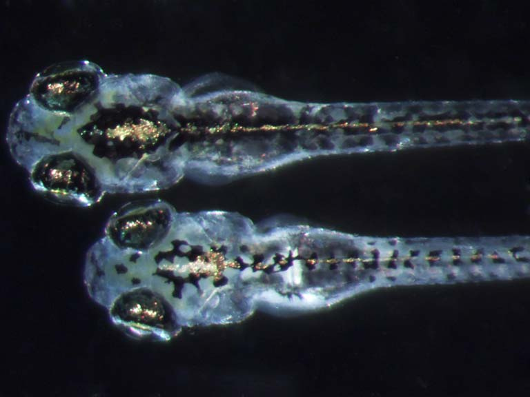
Though humans and fish may seem incredibly different, the zebrafish stripes contain pigmented cells analogous to human melanocytes, melanin containing cells found in eye and skin. Because of these similarities, we were able to construct a zebrafish model of uveal melanoma, a highly metastatic human cancer arising in the eye. The top zebrafish (the bottom fish is a wild type fish control) expresses one of two transgenes harboring mutations detected in 85% of uveal melanoma cases. Expression of the transgene destabilizes the development of the fish melanocytes, thereby giving rise to an aberrant pigment pattern and eventually facilitating tumor formation.
Correct pigment patterning in zebrafish requires proper stem cell differentiation, cell motility and cell- cell signaling. By documenting and understanding the reasons for the change in pigment pattern in the transgene containing fish, we can elucidate the function of the transgene in these pathways.