The Tissue Microenvironment of Unprocessed Human Endometrium
The Tissue Microenvironment of Unprocessed Human Endometrium
Kunzan Liu, Tong Qiu, Jiashu Han, Eva Lendaro, Manuel Levy, Ellen Kan, Keith Isaacson, Fan Wang, Linda Griffith, Sixian You
Koch Institute at MIT, Research Laboratory of Electronics
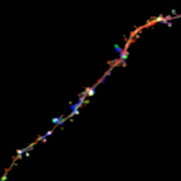
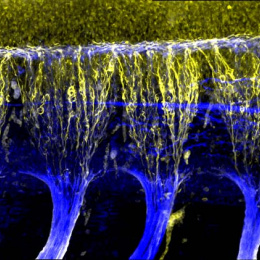
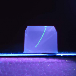
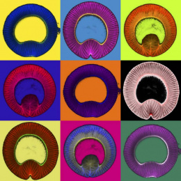
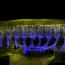
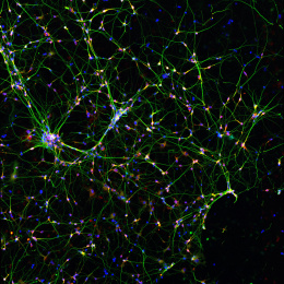
In collaboration with the Wang Lab and Griffith Lab to these series of pictures (3 of 3) were taken to push the limits of label-free optical microscopy, and make it to see deeper, faster, and more structures; and apply the imaging technology to gain more insights into the poorly understood diseases and physiological processes, including the chronic neuropathic pain (whisker pad image) and (endometrium image).
Endometriosis remains a complex and enigmatic disease with no known cure, and the study of endometriosis requires more understandings in the structures and functions of endometrium and its in vitro models. The label-free multicolor imaging of endometrium provides 3D visualizations with sub-cellular resolution that help us to understand its spatial and temporal changes and the imaging of the corresponding in vitro models can monitor the growth of endometrial organoids in real-time non-invasively.

