Tight Embrace of Separating Sisters 2
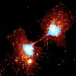
Tight Embrace of Separating Sisters 2
Teemu P. Miettinen, David Mankus, Margaret Bisher, Abigail Lytton-Jean, Scott R. Manalis
Koch Institute at MIT
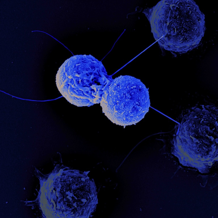
The image displays a leukemia cell undergoing cell division. During this process, a large cell is constricted in the middle of the cell, forming a cleavage that closes until the two resulting sister cells are separated. All the structures seen in this image are folds of the plasma membrane, which is the surface layer of the cell. Our research investigates how these folds form and unfold at different locations on the cell surface as the cell divides, and how this impacts the fidelity of cell division.
