Exploring Cellular Response in Neurons 1
Exploring Cellular Response in Neurons 1
Submitted by Russell McConnell of the Gertler Laboratory at the Koch Institute for Integrative Cancer Research at MIT
MIT Department of Biology, Koch Institute at MIT
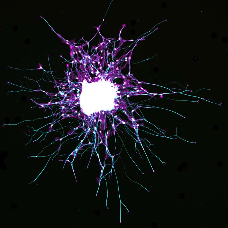
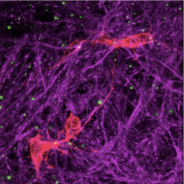
This is an image of spinal cord neurons and supporting glial cells labeled to visualize components of the cells’ skeleton. This type of neuron extends a long protrusion from the spine to sensory receptors in other parts of our body, allowing our brain to receive sensations of touch, pain, and body position. In this image these long protrusions, called axons, are colored in cyan (bluish color) and grow outward from a cluster of neurons in the center (white).
The ability of neurons to sense and respond appropriately to guidance cues is absolutely required during nervous system development and also to repair injuries. Despite the importance of this process, it is not yet known how neurons translate guidance cues into the directed cellular movement that underlies axon guidance. We are seeking to discover how extracellular guidance cues alter the interactions of specific proteins, and how these molecular events produce polarized cellular responses.