Cerebral Organoids Reveal Mitochondrial Defects 4
Cerebral Organoids Reveal Mitochondrial Defects 4
Danielle Tomasello, David Mankus
Koch Institute at MIT, Whitehead Institute
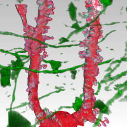
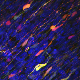
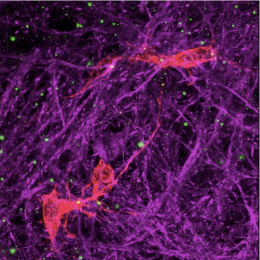
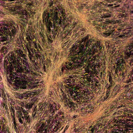
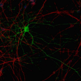
Using human stem cells, we made mini brains to study how Rett Syndrome, a neurodevelopmental disease, develops in the brain. We wanted to look closely at mitochondria to see if there are any differences due to the disease. However, the tissue is very dense, so we tried a method to label different cells in the brain. Neurons were labeled in green and glia were labeled red. We fluorescently imaged the mini brains, then did super-resolution electron microscopy (EM) imaging. After, the fluorescent images were overlayed onto the EM (Correlative Light Electron Microscopy) allowing us to look at the different cell types with high definition.
With this incredibly difficult technique, we were able to identify significant morphological defects in mitochondria between cell types in Rett Syndrome mini brains with high resolution.