Proteomic Human Brain Map
Proteomic Human Brain Map
Juhyuk Park
Picower Institute for Learning and Memory
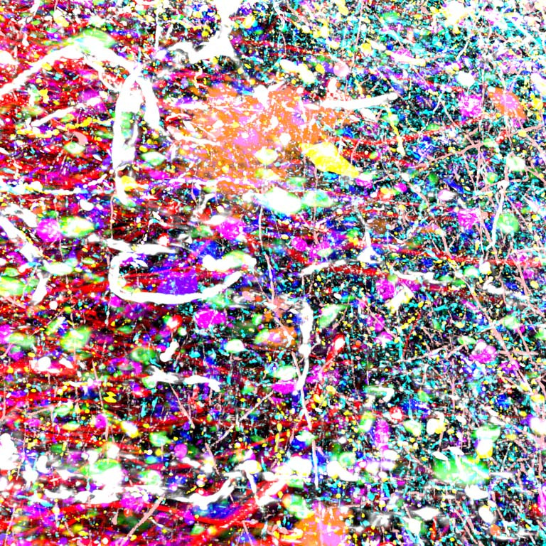
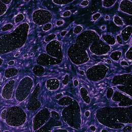
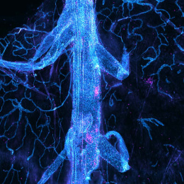
This image shows a complex three-dimensional microenvironment composed of different types of cells in the human brain cortex. It was taken to understand detailed molecular and cellular architectures of brain cells by simultaneously capturing Nuclei (Gray), CR+ neurons (Green), PV+ neurons (Magenta), NPY+ neurons (Yellow), SST+ neurons (Purple), ABETA+ plaques (Orange), GFAP+ astrocytes (Red), IBA1+ microglia (Blue), CD31+ endothelial cells (White), MBP+ oligodendrocytes (Cyan), and SMI-312+ axonal fibers (Pale-pink). We tried to extract spatial, structural, and quantitative information of neuronal and non-neuronal cells in a holistic manner, which is crucial for interrogating human brain function and dysfunction.