Diversity of Immune Cells in the Meninges 1
Diversity of Immune Cells in the Meninges 1
Mitchell Murdock
Picower Institute for Learning and Memory
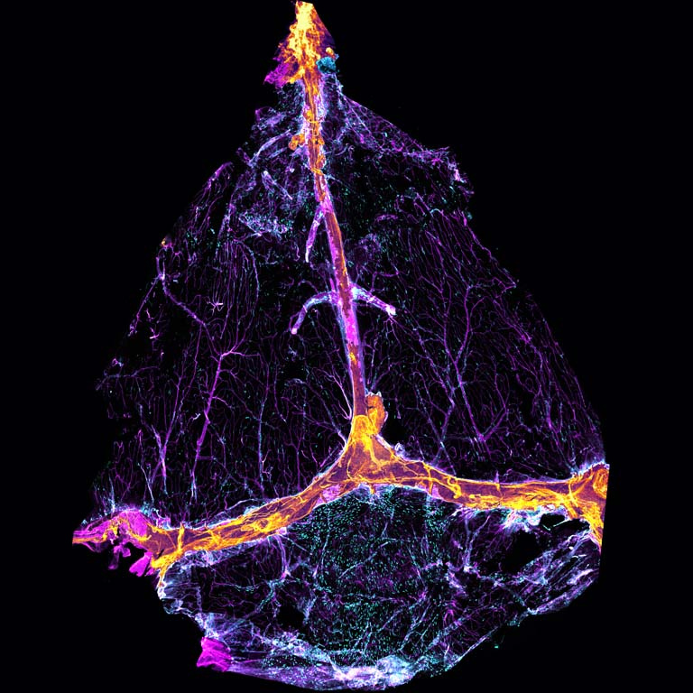
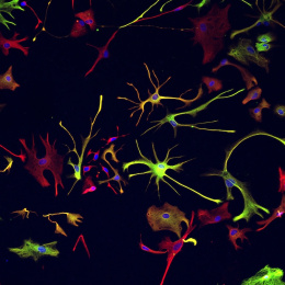
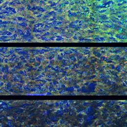
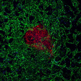
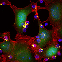
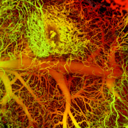
The meninges are a leathery tissue that surround the brain. For decades, pathologists have discarded the meninges as they studied brain anatomy. New evidence from our lab and others suggests that the meninges harbor an astonishing diversity of cells that play a critical role in brain disorders including glioblastoma and Alzheimer’s Disease. These images depict the meninges from a mouse. You can see blood vessels, lymphatic vessels, macrophages (cyan cells in mm1, mm3, and mm4), and even T cells (magenta cells in mm2). Meningeal macrophages actively sense molecules from the brain via cerebrospinal fluid, and other meningeal cells such as T cells—amazingly—secrete substances that enter the brain and modulate neurons, such as specialized interneurons expressing vasoactive intestinal peptide (mm5).
I took these images to visualize (a) whether specific immune cells are present in the meninges, (b) whether/how these cells are disrupted in mouse models of Alzheimer’s Disease, and (c) how these cells respond to patterned light and sound stimulation.