Proof of Principal for Spectral Deconvolution
Proof of Principal for Spectral Deconvolution
Submitted by Matthew Milnes Dobbin of the Tsai Laboratory at the Picower Institute
Picower Institute for Learning and Memory, MIT Department of Brain and Cognitive Sciences
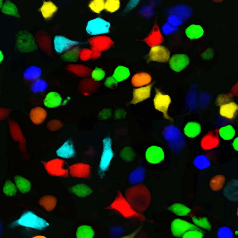
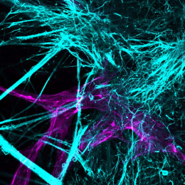
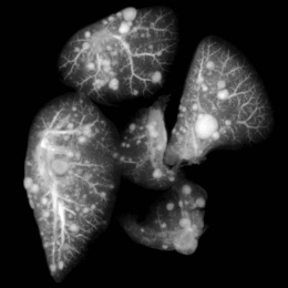
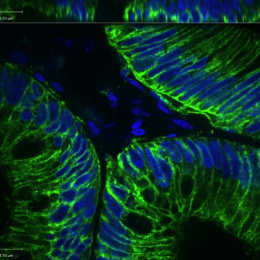
This is an image of a human cell line (293T) in which seven different fluorescent proteins were expressed and subsequently imaged using a specialized imaging technique, spectral deconvolution, that allows for discerning each individual and distinct fluorescent protein color.
The image was generated in the process of my setting up spectral deconvolution on the confocal microscopes in the Tsai Lab, with the ultimate goal of better separating fluorophores with overlapping emission spectra. This image served as a proof of principle for the technique.
Institution of the technique used to generate this image allowed for an overall increasing in flexibility when designing and executing experiments.
