Joint Effort to Repair Cartilage 3
Joint Effort to Repair Cartilage 3
Brett Geiger, Sheryl Wang, Alan J. Grodzinsky, Paula T. Hammond
Koch Institute at MIT, MIT Department of Chemical Engineering
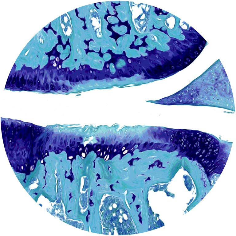
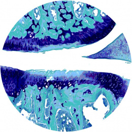
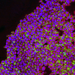
This images shows a cross-section of a knee joint stained with dyes that reveal cartilage damage (top: femur, bottom: tibia, right: meniscus). Joint injuries like an ACL tear accelerate wear and tear on cartilage (blue: healthy, green: damaged), leading to an early onset of painful arthritis. This image is part of a study testing if nanoparticles made in the Hammond lab can enable drugs to penetrate cartilage tissue and repair cartilage.