Expanding the View of the Hippocampus
Expanding the View of the Hippocampus
Fei Chen, Dawen Cai, Ed Boyden
McGovern Institute for Brain Research, MIT Department of Brain and Cognitive Sciences
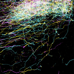
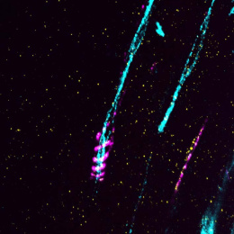
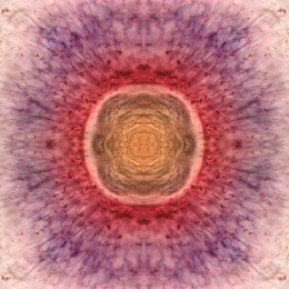
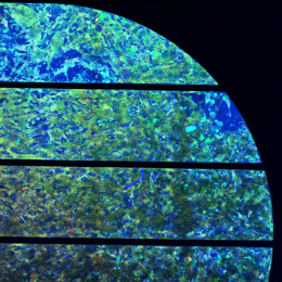
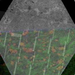
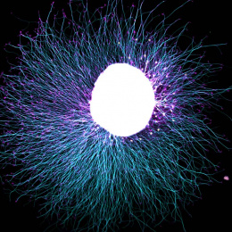
This image is taken of brain section from a mouse— this particular section is called the hippocampus, the area of the brain that is essential for memory and spatial navigation. The surface of different neurons is labeled with combinations of fluorescent colors which allows nearby neurons to be uniquely distinguished. In addition, this particular brain has been physically magnified with a swellable hydrogel, which expands the tissue uniformly in all directions and also makes it transparent in water. This approach—called expansion microscopy—increases the resolving power of optical microscopes, thus letting us look at details which were not visible before under a light microscope. Expansion microscopy enables us to separate closely packed cells in the brain, allowing us to trace their shape and connectivity. Our lab is interested in applying the higher resolutions enabled by expansion microscopy to map circuits in the brain.