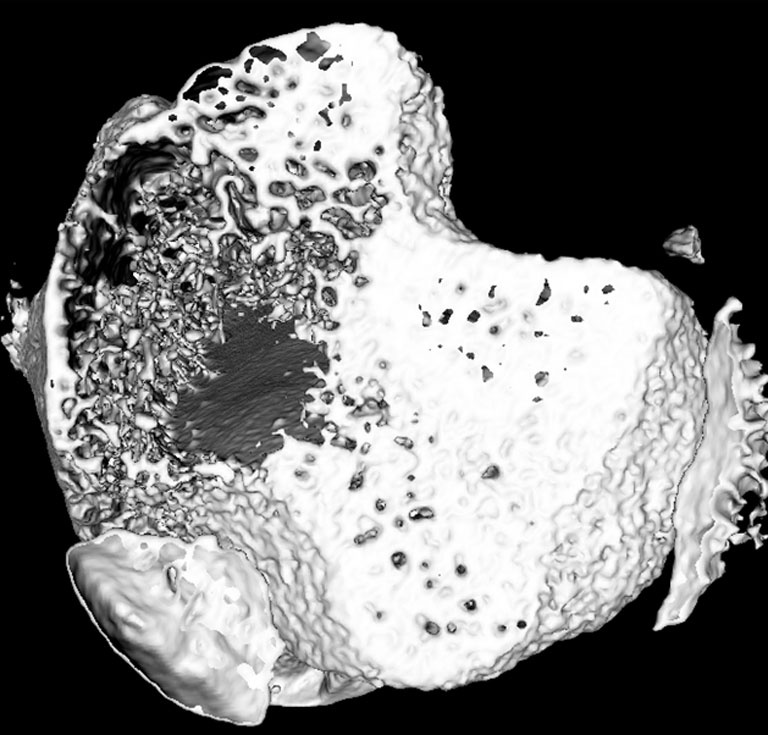
3D Reconstruction of Tibia Bone
3D Reconstruction of Tibia Bone
Submitted by Nisarg Shah of the Koch Institute
Koch Institute at MIT, MIT Department of Chemical Engineering
Nisarg Shah
Hammond Laboratory
Koch Institute
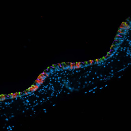
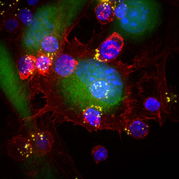
"The fundamental question that we are trying to answer is can we harness the body’s own cells to repair or regenerate human tissue, in cases where it may not be possible without treatment. In this work, we are tailoring nanotherapies that can be used to direct cells to for bone tissue. The images submitted here depict the living bone tissue environment at the microscopic level that is highly dynamic.
This image shows a 3D reconstruction of an entire segment of the tibia bone, taken using micro-computed tomography (μCT) scans, that are commonly used in clinical settings to identify abnormalities in the tissue. The image depicts the organization of the bone just below the knee joint, which is composed of trabecular bone (plate like spicules) on the inside and a continuous dense core on the outside, which is cortical bone. The structure is perpendicular (coming out) of the plane of the image (i.e. you are looking down the bone canal)."