Transparent Neuron: A Rendering
Transparent Neuron: A Rendering
Submitted by Daniel Berger and Sebastian Seung (MIT), and Narayanan 'Bobby' Kasthuri, Ken Hayworth, Richard Schalek, Juan-Carlos Tapia and Jeff Lichtman (Harvard)
MIT Department of Brain and Cognitive Sciences, Harvard University Department of Molecular and Cellular Biology
Daniel Berger and Sebastian Seung
MIT Department of Brain & Cognitive Science
Narayanan 'Bobby' Kasthuri, Ken Hayworth, Richard Schalek, Juan-Carlos Tapia and Jeff Lichtman
Harvard University Department of Molecular and Cellular Biology
Electron Microscopy and 3D Modeling
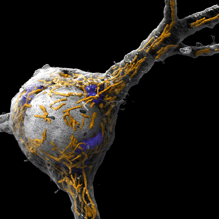
"Our images show 3d renderings of neurons and parts of neurons which were reconstructed from electron-microscopic images of a very large stack of slices of mouse somatosensory cortex. This particular image shows the cell body of a neuron in mouse somatosensory cortex, with its nucleus (white), mitochondria (orange), Golgi apparatus (blue) and lysosomes (black) labeled. The endoplasmic reticulum is not shown. Labeling the organelles of a cell enables us to better understand their shape and spatial distribution within the cell. The colors are all arbitrary since the EM images do not show color."