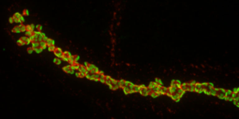
A Motor Neuron Synapses Onto a Muscle, Version #1
A Motor Neuron Synapses Onto a Muscle, Version #1
Submitted by Jan Melom, Picower Institute for Learning and Memory
Picower Institute for Learning and Memory, MIT Department of Brain and Cognitive Sciences
Jan Melom
Picower Institute for Learning and Memory
Spinning Disc Confocal Microscopy
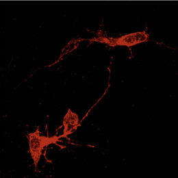
"Our lab is interested in studying molecular mechanism of synaptic transmission and morphology. This image shows a Drosophila motor neuron synapsing onto a muscle (in the plane of the screen). It was taken while studying a knockout animal disrupted in NHE3, a gene recently linked to autism spectrum disorders that we are studying in the lab. The surface of the neuron is colored red. In green are glutamate receptors expressed on the muscle that detect neurotransmitter release from the neuron. The resolution of the image allows you to see individual sites of synaptic vesicle release and detection peppering the surface of the neuromuscular junction."