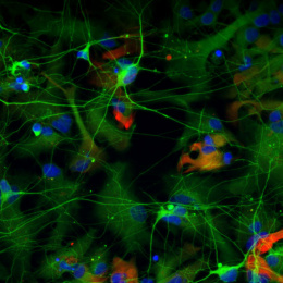
Long Term Growth and Maturation of Neurons Grown in a Microplate
Long Term Growth and Maturation of Neurons Grown in a Microplate
Submitted by Asha L. Bhakar and Mark F. Bear of the Picower Institute for Learning and Memory
Picower Institute for Learning and Memory, MIT Department of Brain and Cognitive Sciences
Asha L. Bhakar and Mark F. Bear
Picower Institute for Learning and Memory
Epi-Fluorescence Micrograph
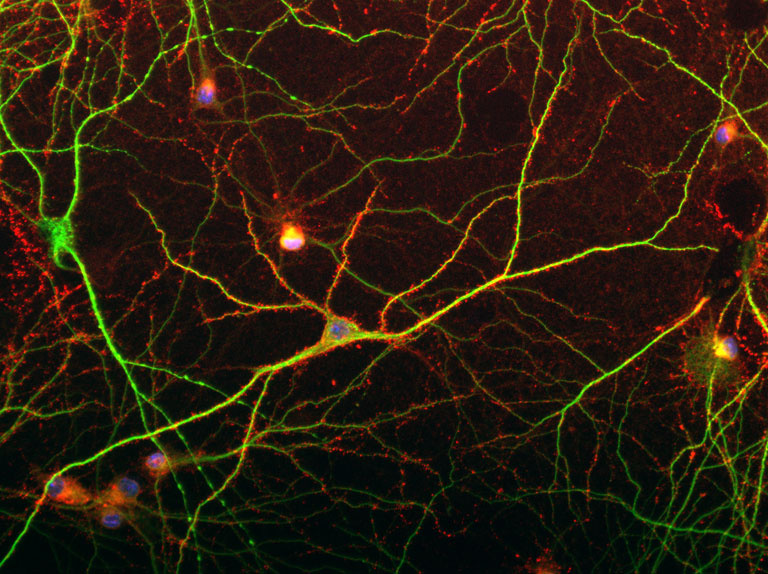
"This image is part of the work we are doing to develop a high-throughput microplate assay that specifically studies synaptic connectivity defects believed to underlie cognitive impairment in autism spectrum disorders (ASDs), and then use this assay to discover new biomarkers and treatments for the disease. In the image, dissociated hippocampal neurons grown for 21 days in optical 96-well plates show features of neuronal maturation including pyramidal shaped somas, extensive dendritic arbors (green), and discrete punctate appearance of the post-synaptic protein PSD-95 (red) along the dendrites. Nuclei are labeled with Hoeschst-33342 (blue)."
