Ignition Sequence 2
Ignition Sequence 2
Liz Fink, William Pinney, and K. Dane Wittrup
Koch Institute at MIT, MIT Department of Biological Engineering
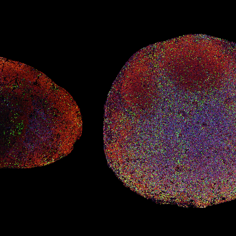
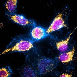
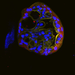
Down-sampled from multi-billion-pixel raw data, this image captures the ignition of a potent anti-cancer immune response in single-cell resolution. Both tissue sections come from mice bearing identical aggressive melanoma tumors, yet these animals face completely divergent fates.
The lymph node on the left belongs to an untreated mouse that will succumb to its tumors within two weeks; the node on the right belongs to a mouse treated with a triple combination of precisely engineered immune-stimulatory cytokines developed at the Koch Institute, shown to trigger complete tumor regression. The difference is impossible to miss: the treated node has swollen to many times its original size, engorged by a vast influx of new immune cells.
Orange nuclei reveal the hidden architecture of each node; red halos mark antigen-presenting cells primed to educate T cells against the tumor; blue fills the T cell zone — vastly expanded in the treated node as activated T cells proliferate thousands of times over. The blazing green and yellow spots are cells caught mid-division, clustering across the treated tissue in pockets of frantic activity. This image captures not just cellular behavior, but the difference between life and death.