Koch Institute Image Awards Archive

The Koch Institute Image Awards celebrate the extraordinary visuals produced through life sciences and biomedical research at MIT.

Primary Nav

  • Explore images
    • View by collection
    • View by keyword
    • Surprise me!
  • Learn more
    • KI Image Awards
    • Support us
    • Visit
    • Image use
  • View images by keyword
  • Surprise me!
Koch Institute

Blue Spleenies 

Blue Spleenies 

Punya Gupta, Tyler Jacks

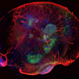
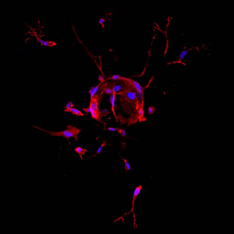
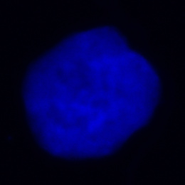
This is a cross-sectional image of a spleen from a mouse, in which we can see clearly visualize the spatial distribution of different cell types.

We can identify a prominent macrophage population and distinct T cell zones in the spleen of our murine model.

Light blue rounded shapes surrounded by grey rough terrain

More like this

Fluorescent pink landscape covered over with fiery purple clusters.
Cyan strands, dotted in white, organize in a spiral, curved clockwise from center. After 1.5 loops the spiral heads upward and the cyan ends at magenta and yellow spots before reaching a rounded point with clustered white dots.
White webbing and globule resembling snow littering a pale periwinkle backdrop.
a central mass of brain tissue with arc-like tissue structures around it

Keywords

  • 2025
  • cell differentiation
  • cell proliferation
  • confocal microscopy
  • Jacks Laboratory
  • macrophages

Koch Institute for Integrative Cancer Research
Massachusetts Institute of Technology
500 Main Street, Building 76
Cambridge, MA 02139-4307

  • Contact
  • Image Use
  • Accessibility
  • Visit us on Facebook
  • Visit us on X
  • Visit us on LinkedIn

© 2025 Koch Institute for Integrative Cancer Research