Lymph Bizkit, 2
Lymph Bizkit, 2
Anna Romanov, Mark Bathe, Darrell Irvine
Koch Institute at MIT
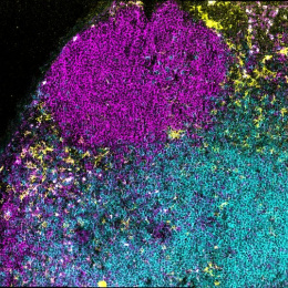
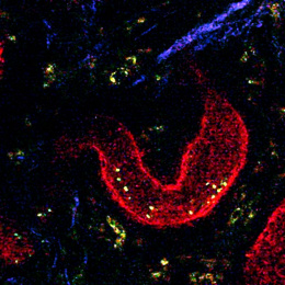
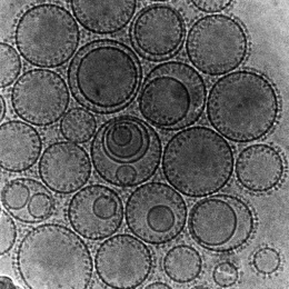
These microscopy images show what happens in draining lymph nodes shortly after vaccination. Vaccine antigens (red) are scavenged by defensive cells called subcapsular macrophages (green). Depending on the antigen formulation, the antigens may be retained in the green macrophages or be handed off to follicular dendritic cells (blue). This “relay” mechanism determines the magnitude of immune response after vaccination.
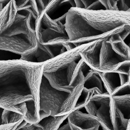
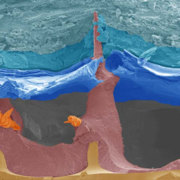
These images elucidate different trafficking behaviors of vaccine nanoparticles in lymph nodes as well as show the sophisticated filtration system of the lymph node and the elaborate immune hub that is at work during vaccination.