The Ghost of a Flea, 2
The Ghost of a Flea, 2
Eric Nguyen, Kaelan Reed, Arnab Rudra, Akash Gupta
Koch Institute at MIT
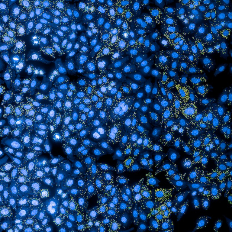
For mRNA vaccines to work, the mRNA has to be released from tiny nanoparticles into the cell. This image is from an assay we use to screen nanoparticles to make better mRNA vaccines. In this assay, we dose cells with mRNA vaccine formulations and then use a technique called single molecule fluorescence in situ hybridization (smFISH) to light up the mRNA inside the cells. This lets us see all of the mRNA is inside the cells, and how much of it is still trapped in nanoparticles or free.
In the picture, the solid green spots show mRNAs that are still stuck inside the nanoparticles. On the other hand, the yellow spots show mRNA that has successfully escaped from the nanoparticles into the cell, where it can initiate a vaccine response.
We took this image to evaluate how effectively different lipid nanoparticle (LNP) formulations deliver mRNA into cells and facilitate its escape from the endosome.
The most exciting part of this image is that it shows how our lipid nanoparticle formulation outperforms some commercial LNPs, potentially leading to more effective and safer mRNA therapies with lower doses and reduced toxicity.