Dance of the Conjoined Sister Cells
Dance of the Conjoined Sister Cells
Teemu P. Miettinen and Scott R. Manalis
Koch Institute at MIT
The act of cell division requires a systematic reorganization of a cell to ensure that various cellular components can be appropriately partitioned to the two daughter cells. First, a cell pulls apart the two copies of its DNA. This is followed by a constriction of the cell in the middle to separate the rest of cellular contents into two, until only a small bridge connects the two daughter cells. However, sometimes this process makes mistakes, which can alter the contents and behavior of the daughter cells and result in cancer initiation and progression. Knowledge about how a cell divides and how the process of cell division can fail, will help us understand cancer and find cancer specific vulnerabilities that may enable novel cancer treatments.
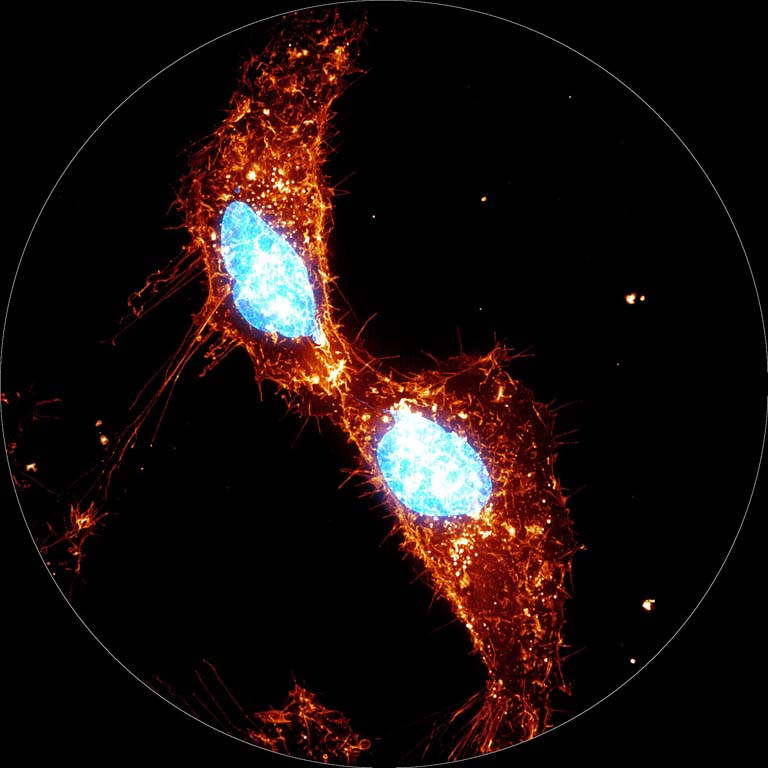
Work in the Koch Institute, led by Dr Miettinen and Prof Manalis, has investigated the dynamics of the cell’s outer membrane, also known as plasma membrane, during cell division. The image displays two live human adenocarcinoma sister cells, which have just divided from a mother cell. The plasma membrane of the cells is labelled with red and yellow colors. Many membrane protrusions can be seen that help the cell attach and move around its environment. The shown sister cells are still connected to each other through a thin intercellular bridge, within which a narrow strand of DNA, labelled in blue, can be seen. This DNA strand is known as a chromatin bridge, and it represents the cell’s failure to segregate the DNA between the daughter cells normally. Such chromatin bridges can result in DNA breakage and mutations that drive cancer. By understanding how the plasma membrane movement is regulated during cell division, the researchers hope to explain how such cell division errors arise and how they can be corrected.
