Chromatin Loop Extrusion 2
Chromatin Loop Extrusion 2
Michele Gabriele
MIT Department of Biological Engineering
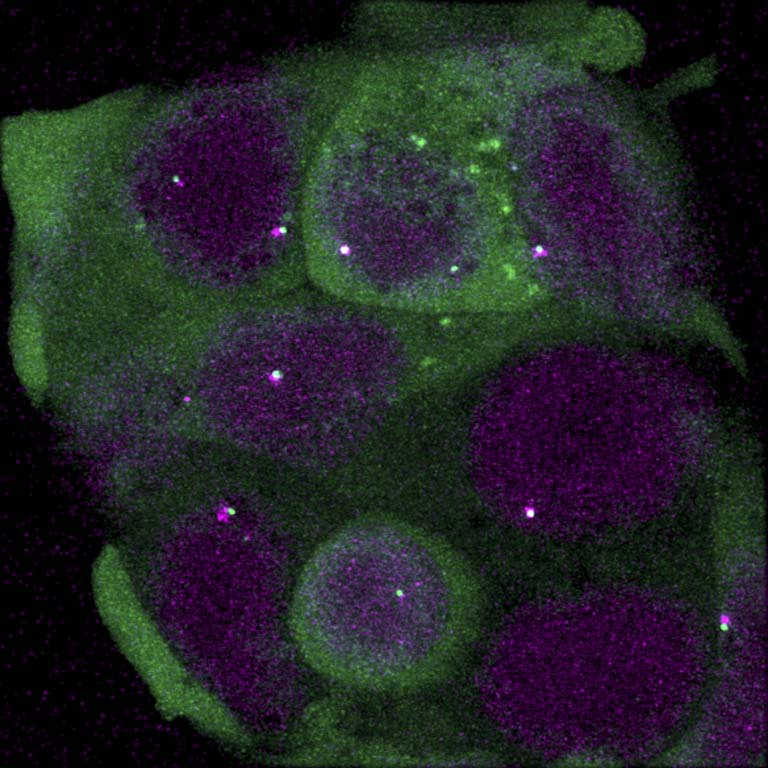
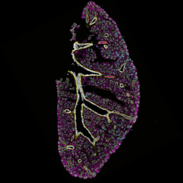
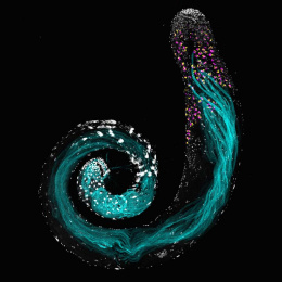
These pictures represent living mammalian cell in which we can distinguish the cytoplasm (green) and the nuclei (purple), where the DNA is located. The bright purple and green dots inside the nuclei are two labels to identify two distant regions of DNA located in the same DNA chain. What makes unique these pictures is that the cells are alive and we can measure the dynamics of the contacts of these two labeled DNA regions to understand the phenomenon of chromatin organization. This is an essential biological process that regulates development.
We record movies of chromatin dynamics to understand how the chromatin is folded inside nuclei. With these experiments we try to prove the existence of chromatin loop extrusion in vivo and we try to measure its dynamics and duration in living cells. These images are part of a real larger collection of movies used for data analysis.