LSECs Deform a Hydrogel 2
LSECs Deform a Hydrogel 2
Alex Wang, Allysa Allen
MIT Department of Biological Engineering, Koch Institute at MIT
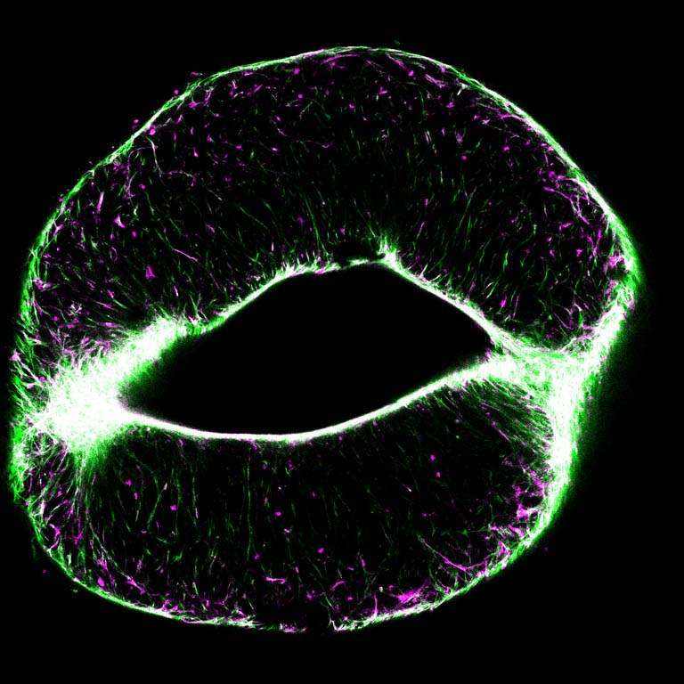
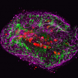
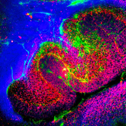
A major challenge in the field of organoid engineering is creating vascularized tissue constructs. Our larger research goal is to build in vitro models of vascularized liver (and other epithelial cells) in synthetic, reproducible, and well-defined environments. Doing so will allow us to better model the full range of biological responses and probe complex liver biology.
This image depicts human liver sinusoidal endothelial cells (LSECs) encapsulated in a synthetic hydrogel made of large polyethylene glycol (PEG) molecules. PEG hydrogels, with their tunable chemistry and material properties, can be designed to mimic a biological matrix. When encapsulated into our engineered PEG hydrogels, LSECs form a vascular network-like structure. They also severely deform and contract the gel, ripping it apart in the center. The once fully circular gel is compacted to approximately half its size, and its middle has been squeezed out. The colors depict cell cytoskeletal f-actin (green) and endothelial surface marker CD31 (magenta). The high concentration of f-actin in the center shows how the cells are able to exert enough force to rip open the soft gel. [This] image is a single 50um slice of the gel.