Imaging A Zebrafish Brain
Imaging A Zebrafish Brain
Danielle Tomasello
McGovern Institute for Brain Research, MIT Department of Brain and Cognitive Sciences, MIT Department of Biology, Whitehead Institute
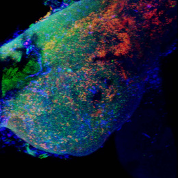
This is an image of an adolescent zebrafish brain. We are comparing normal to mutant brains. Mutant brains showed ectopic staining of a synaptic protein. These experiments help us understand what the developing brain should look like and how our mutant brains differ. The SHIELD preservation and clearing of the brain, not yet performed on zebrafish tissue, keeps tissue beautifully intact and allows imaging of whole brains without need of sectioning.

