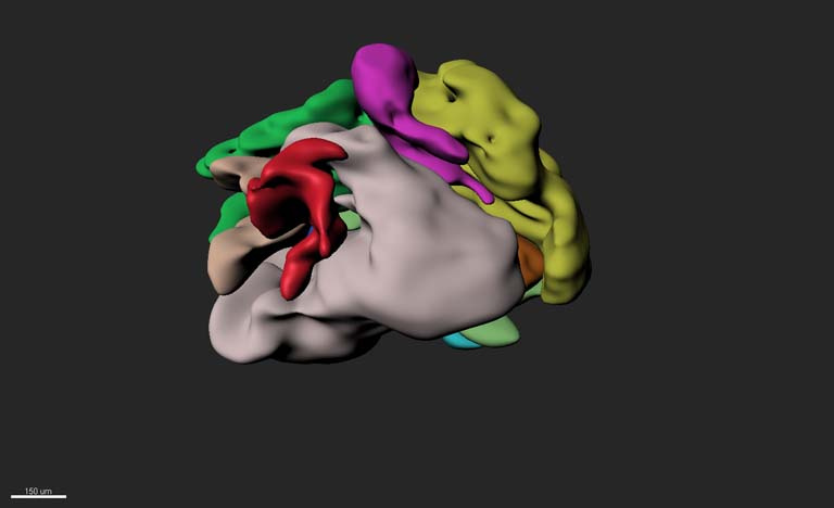
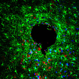
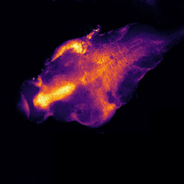
Ventricular Zones in a Cerebral Organoid
Ventricular Zones in a Cerebral Organoid
Murat Yildirim
Picower Institute for Learning and Memory, MIT Department of Brain and Cognitive Sciences
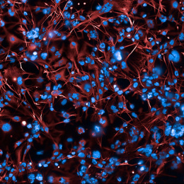
Studying neurodevelopmental disorders with animal models is challenging due to the fact that the stages of brain development in animal models and human are different. Thus, there is an important requirement to model these neurodevelopmental disorders in the lab with human cells. In our lab, we are generating cerebral organoids, in other words mini brains, from the stem cells that we acquire from patients’ skin in order to recapitulate early events of embryonic brain development. Rett Syndrome is a neurodevelopmental disorder that affects only girls. Its symptoms are most often reported to emerge between 6 and 18 months of age. Therefore, it is important to understand fundamental mechanisms to cause this disease and develop therapeutic strategies to fight against this disorder.
Since this is an early stage neurodevelopmental disorder, the abnormalities more likely occur in the early stages of brain development. In developing human brain, there are several ventricle-like cavities (ventricular zones) where neurons move outwards to generate the compartments of the brain. Therefore, it is crucial to determine whether there are any abnormalities in these ventricular zones in the developing brain. In order to answer this question, we capture three-dimensional picture of these cerebral organoids derived from Rett Syndrome patients with the home-made microscope that I built at MIT. In this image, each color represents a different ventricular zone in three-dimensional space and the shapes of these ventricular zones are too different than uniform and spherical shape which is observed in healthy cerebral organoids.