Investigating Alzheimer's Disease in the Visual Cortex 2
Investigating Alzheimer's Disease in the Visual Cortex 2
Mitchell Murdock
Picower Institute for Learning and Memory, MIT Department of Brain and Cognitive Sciences
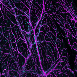
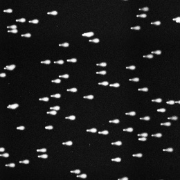
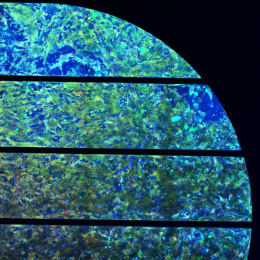
These images are montages of the vasculature of the living mouse visual cortex. I installed a cranial window over the primary visual cortex of a mouse engineered to recapitulate Alzheimer’s pathology.
As you look from left to right and top to bottom, each frame corresponds to 1 micron imaged deeper into the cortex. You can trace minute capillaries (the smaller vessels) as well as large arterioles plunging deep into the brain (bright circles perpendicular to the imaging plane). If you look carefully at some of the capillaries, you can see tiny shadows—these correspond to individual red blood cells and immune cells which do not take up the dye and therefore appear as dark patches against the bright plasma. During the time lag of each optical plane adjustment, you can even see cells move along the capillary. In some capillaries, the blood cells do not move, indicating that this is a “plugged” capillary, thought to occur because the cells adhere to the inflamed capillary wall.