A Cellular Network Assembles Within the Heart 2
A Cellular Network Assembles Within the Heart 2
Alexander Auld and Laurie Boyer
MIT Department of Biology, MIT Department of Biological Engineering, Koch Institute at MIT
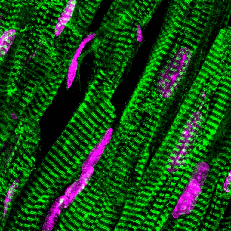
These pictures are of cardiomyocytes, which are heart muscle cells. They contain a highly organized contractile network (green), called the sarcomere, and nuclei (magenta). These cells are responsible for pumping blood throughout the body for the duration of life. Our lab is trying to understand how cell metabolism impacts the formation of the contractile network of the cardiomyocyte during development.
This image was taken from a slices of adult mouse heart tissue. For heart tissue we modified a relatively new technique called expansion microscopy. Expansion microscopy involves embedding the tissue within a hydrogel and then swelling the gel, which expands the tissue. The increase in the size of the tissue allows for super resolution images to be taken on diffraction limited microscopes. Cardiac troponin is colored in green to visualize the sarcomere and nuclei in magenta.