Mechanical Properties of the Cell: Studying Stiffness
Mechanical Properties of the Cell: Studying Stiffness
Teemu Miettinen
MIT Department of Biological Engineering, Koch Institute at MIT
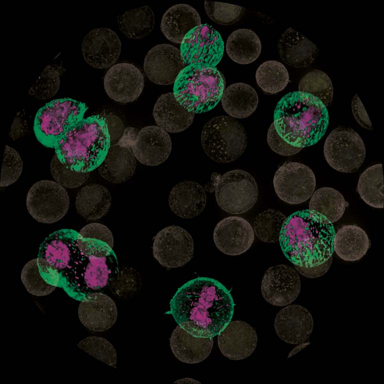
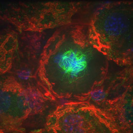
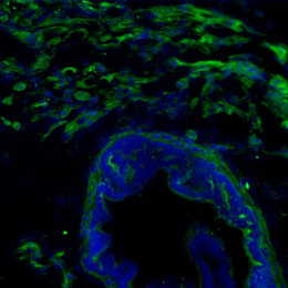
This image displays live mouse blood cancer cells in different stages of their growth and division cycle. These cells grow in suspension, not adhering to their environment, which shows as a round cell shape, and it also makes the study of these cells challenging. Starting from the top right and moving clockwise, a small cell grows in size, duplicates its’ cellular components, such as DNA (pink color), and divides in to two separate daughter cells. These different stages, especially the cell division, have to be coupled to the control of cell’s outer support structure called actin cortex (green color). Underlying the individual cell pictures, one can see a population of these cells (actin cortex is displayed in black&white and DNA is displayed in yellow). When we combine imaging such as this to other measurements developed here at MIT, we can study how the structure of the actin cortex affects the surface stiffness of a cell. This will help us understand, for example, how cancer cells are able to change their mechanical properties and invade new tissues
