Tissue Regeneration after a Stroke Injury 1
Tissue Regeneration after a Stroke Injury 1
Submitted by Dr. Myron Spector (Senior Lecturer, Department of Mechanical Engineering) and Dr. Teck Chuan Lim (Post-doctoral Fellow, Tissue Engineering Laboratory)
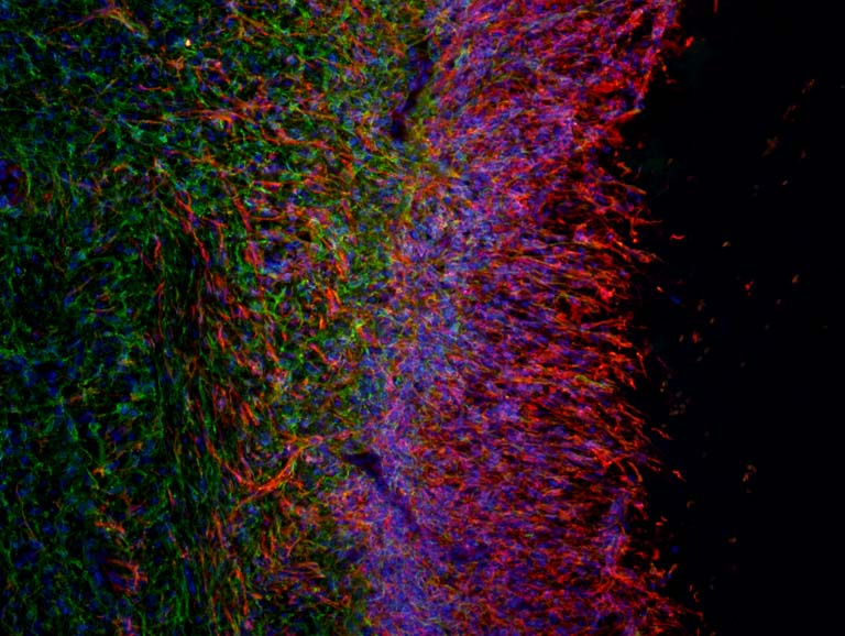
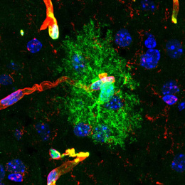
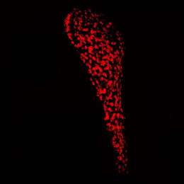
The image depicts a zone of tissue regeneration within a rat brain after stroke injury. Mature mammalian brains are not known to regenerate spontaneously after injury. To overcome this challenge, an injectable matrix containing epidermal growth factor was engineered and injected into the injured region. Given the stimulus and structural support provided by the bioactive matrix, neural stem and progenitor cells (marked red for their expression of nestin) grew extensively beyond the original brain tissue discerned by the brain supporting cells, astrocytes (marked green for their expression of glial fibrillary acidic protein) into the implanted matrix.
The image was taken for the purposes of: 1) determine the identity of cells within neotissues extending into the implanted matrix and 2) investigate the expression of nestin (neural stem and progenitor cell marker) in relation to that of glial fibrillary acidic protein (GFAP; astrocyte marker).
