Viewing Lung Tumors in 3D, Image 3
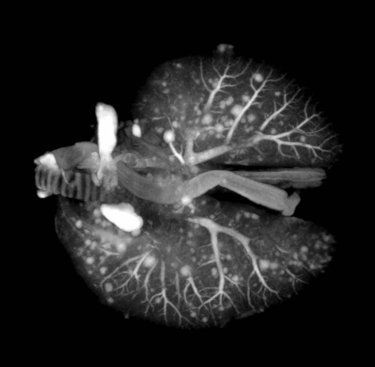
Viewing Lung Tumors in 3D, Image 3
Submitted by Milton Cornwall-Brady of the Applied Therapeutics and Whole Animal Imaging Core at the Koch Institute
Koch Institute at MIT
"Mouse lungs were scanned in a microCT, generating a 3D dataset that allows researchers to count the number of tumors and measure their size. These images help us to understand the distribution of tumors within the lungs as well as quantifying any changes in the number/size of tumors due to whatever form of intervention (e.g., chemotherapy) is being tested. Although this can be done with traditional histology, with this method we obtain a full 3D measurement of all tumors present, not just a 2D slice through a random subset of tumors as one would get from histology."
