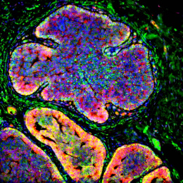
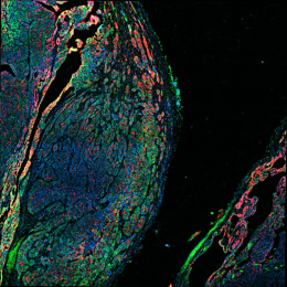
Metastatic Breast Cancer Invades a Spleen
Metastatic Breast Cancer Invades a Spleen
Submitted by Alexandra Naba of the Hynes Laboratory at the Koch Institute.
MIT Department of Biology, Koch Institute at MIT
Alexandra Naba
Hynes Laboratory, Koch Institute
Light Micrograph
"This image shows a section of the spleen of a mouse bearing a metastatic mammary tumor. Two metastases are visible in the image and appear bright pink within the normal splenic tissue (darker pink/purple).
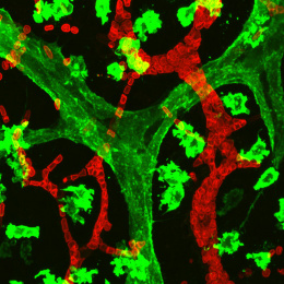
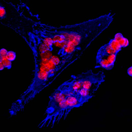
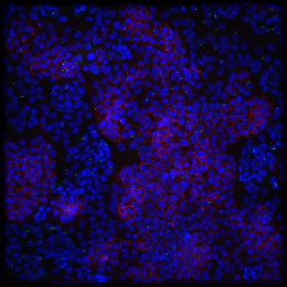
I am interested in understanding how the extracellular matrix influences tumor progression and metastasis formation. The extracellular matrix constitutes the architectural scaffold that supports cells within tissues. Alterations in its organization and/or changes in its composition have been shown to promote tumor progression. Collagens – depicted in the images submitted – are one of the main components of the extracellular matrix and I use it as a marker of tumor progression, as the level of extracellular matrix deposition and organization correlates positively with a more advanced stage of tumor progression. My research goal is to characterize the changes in the composition of the extracellular matrix during tumor progression in order to identify novel biomarkers that will serve as prognostic and diagnostic tools for patients with cancer."