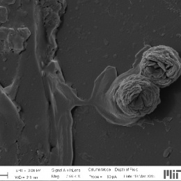
Metastatic Breast Cancer Invades a Liver, Version #2
Metastatic Breast Cancer Invades a Liver, Version #2
MIT Department of Biology, Koch Institute at MIT
Alexandra Naba
Hynes Laboratory, Koch Institute
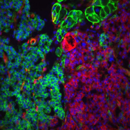
Light Micrograph
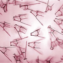
"This image shows a section of the liver of a mouse bearing a metastatic mammary tumor. At the center of the image is a metastasis coming from the mammary tumor. The normal hepatic tissue appears in pink/purple and surrounds the metastasis at the center of the image. The characteristic of the metastasis is the dramatic deposition of collagen fibers (in blue) within and around it. Moreover, one can note that the metastasis is not a well-defined secondary tumor but emits strands of cells in the normal hepatic tissue, indicating the invasive nature of the metastatic cells.
I am interested in understanding how the extracellular matrix influences tumor progression and metastasis formation. The extracellular matrix constitutes the architectural scaffold that supports cells within tissues. Alterations in its organization and/or changes in its composition have been shown to promote tumor progression. Collagens – depicted in the images submitted – are one of the main components of the extracellular matrix and I use it as a marker of tumor progression, as the level of extracellular matrix deposition and organization correlates positively with a more advanced stage of tumor progression. My research goal is to characterize the changes in the composition of the extracellular matrix during tumor progression in order to identify novel biomarkers that will serve as prognostic and diagnostic tools for patients with cancer."