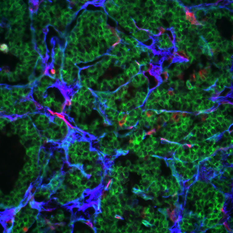
Deposition of Fibronectin Around a Tumor
Deposition of Fibronectin Around a Tumor
Submitted by Patrick Murphy, Hynes Lab, Koch Institute
MIT Department of Biology, Koch Institute at MIT
Patrick Murphy
Hynes Lab, Koch Institute
Confocal Microscopy
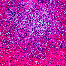
"The image shows the deposition of fibronectin (blue) around the vasculature in a growing spontaneous pancreatic tumor. To determine the utility of a specific deletion strategy in the tumor, I used a mouse line in which Cre activity causes a switch in expression from membrane bound green-fluorescent protein to membrane bound red-fluorescent protein. This image is from my control, an animal in which the reporter and Cre were present, but the floxed fibronectin allele was not. Here, the cells of the tumor have switched from red to green, but fibronectin expression remains. In the mutant, cells have switched and fibronectin expression is lost."