Young Neurons Mature in a Microplate
Young Neurons Mature in a Microplate
Submitted by Asha L. Bhakar and Mark F. Bear of the Picower Institute for Learning and Memory
Picower Institute for Learning and Memory, MIT Department of Brain and Cognitive Sciences
Asha L. Bhakar and Mark F. Bear
Picower Institute of Learning and Memory
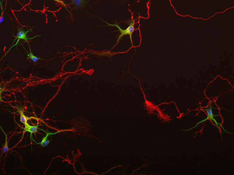
Epi-Fluorescence Micrograph
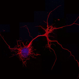
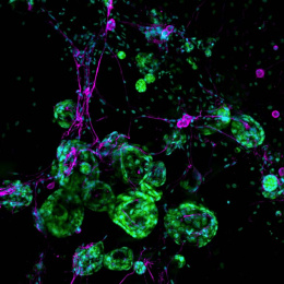
"This image was taken from a series of optimization experiments, designed to determine how best to miniaturize, visualize and analyze synaptic and dendritic structures in primary neurons grown in optical 96-well plate formats and imaged by automated epi-fluorescent microscopy. In the image, seven day-old dissociated hippocampal neurons show simple, immature dendritic arbors (green) extending from neuronal cell bodies together with long pathfinding axons with large growth cone heads (red) when grown in optical 96-well plates."
