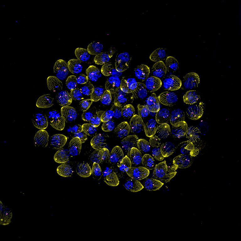
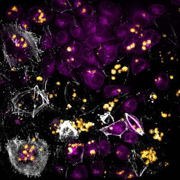
Disrupted Division
Disrupted Division
Dominic Schwarz, Benicio Tapia, Sebastian Lourido
Koch Institute at MIT, Whitehead Institute
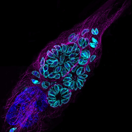
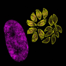
This ultrastructure expansion microscopy image shows a parasitophorous vacuole filled with Toxoplasma gondii parasites at the onset of cell cycle disruption. Parasite microtubules are shown in yellow, DNA in blue, and centrosomes marked by Centrin in magenta. These parasites lack TgBRM, a chromatin remodeler that maintains transcriptional readiness during cell cycle progression. Subtle abnormalities—including parasites with too many or too few centrosomes—hint at the breakdown in division control that ultimately leads to multinucleated cells.