Ignition Sequence 3
Ignition Sequence 3
Liz Fink, William Pinney, and K. Dane Wittrup
Koch Institute at MIT, MIT Department of Biological Engineering
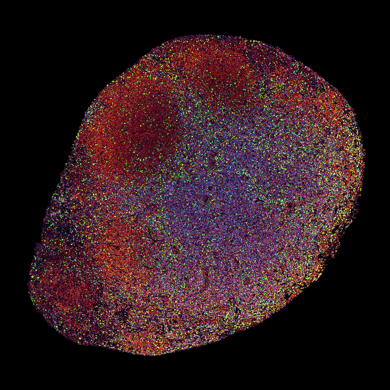
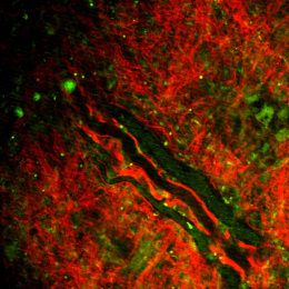
Down-sampled from multi-billion-pixel raw data, this image captures the ignition of a potent anti-cancer immune response in single-cell resolution. This tissue section comes from a mouse bearing an aggressive melanoma tumor, treated with a triple combination of precisely engineered immune-stimulatory cytokines — a therapy shown to trigger complete tumor regression.
What we are seeing is a lymph node in the midst of that transformation: swollen to many times its original size, engorged by a vast influx of new immune cells as the immune system springs violently into action. Orange nuclei reveal the hidden architecture of the node; the red halos surrounding many cells mark antigen-presenting cells, loaded with material from dying tumor cells and primed to educate T cells on how to recognize this foe. Through the center, shades of blue mark the T cell zone — vastly expanded as activated T cells proliferate thousands of times over. The blazing green and yellow spots are cells caught mid-division, many still visibly elongated, clustering across the tissue in pockets of frantic activity. This is immunity at full throttle — a chain reaction, ignited.