Comb without the Honey
Comb without the Honey
Vardhman Kumar, Joa Yun, Heather Fleming, Sangeeta Bhatia
Koch Institute at MIT
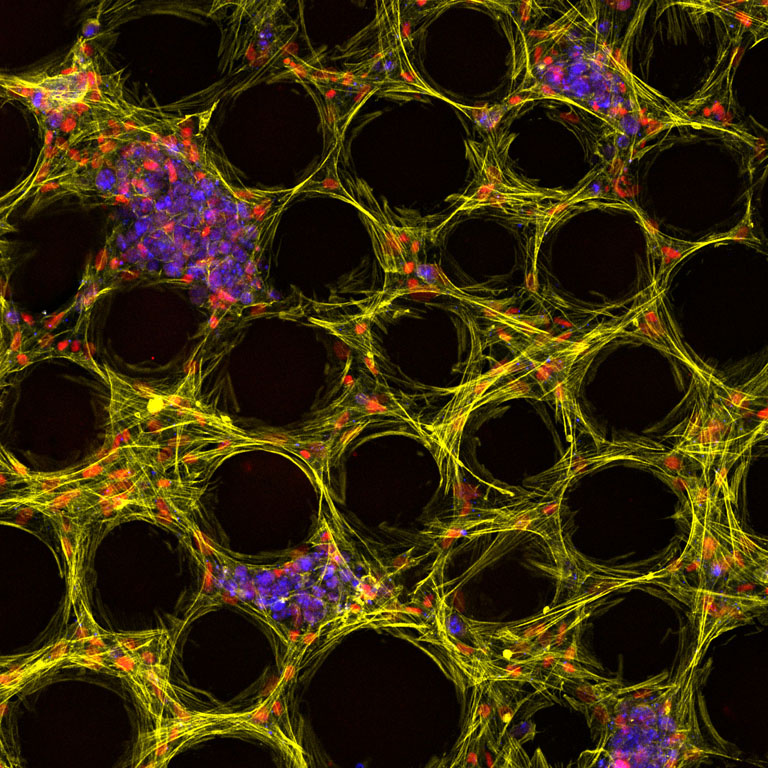
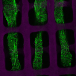
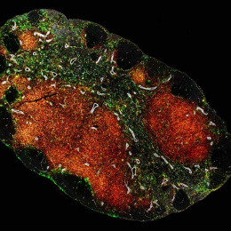
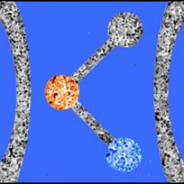
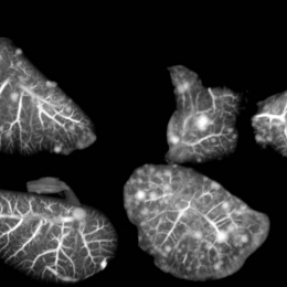
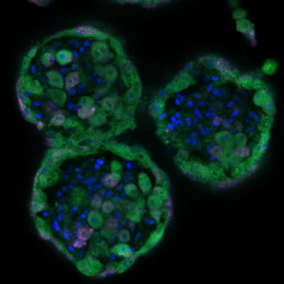
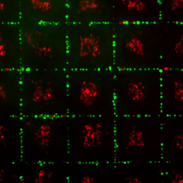
This image illustrates a 3D liver tissue construct consisting of hepatocytes (cyan), and supporting fibroblasts (red) and nuclei (yellow). To have the observers focus on the negative space, we have omitted the microspheres in this image. The hydrogel provides a matrix that mimics the liver’s extracellular environment, organizing cells into functional architectures and enabling nutrient exchange. Notice the cells in the spaces between the green microspheres, the magenta hepatocytes clustering along the tiny corridors and the yellow fibroblasts lining and remodeling these channels. Together, these components create a bioengineered liver microenvironment that can model tumor–stroma interactions and potentially bridge the gap between cancer therapy and regenerative medicine for patients awaiting transplantation.