Fluorescence and the Machine
Fluorescence and the Machine
Shixun Han, Fangtao Chi, Omer Yilmaz
Koch Institute at MIT
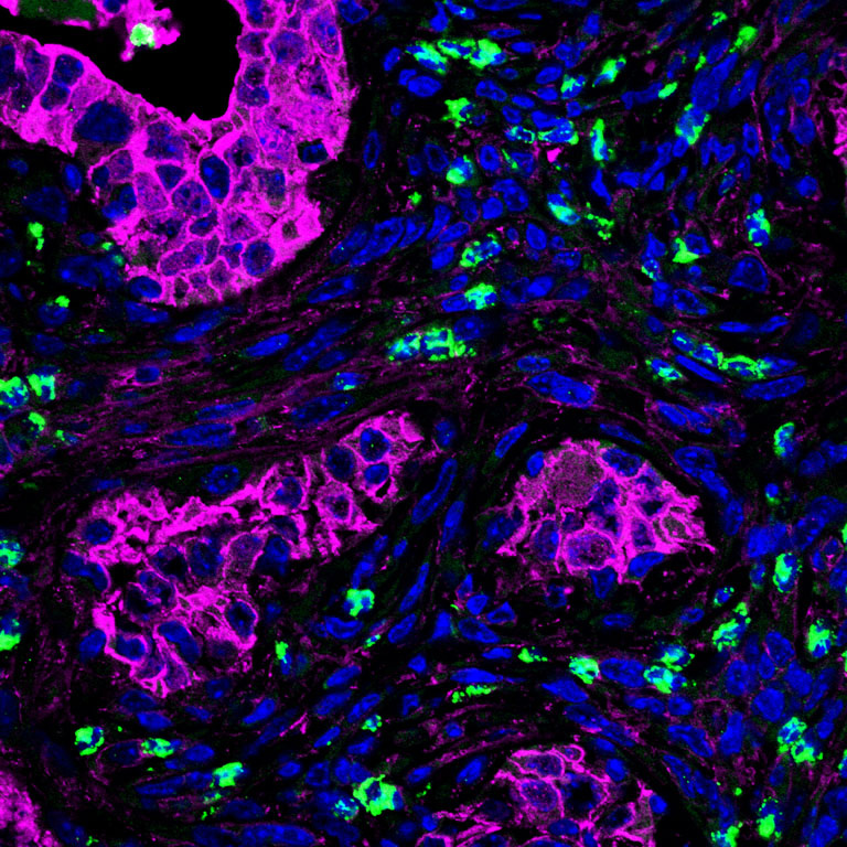
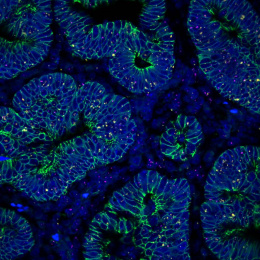
This image captures a mouse colon tumor generated by colonoscopic injection of APC-deficient organoids, visualized through fluorescent imaging. The red fluorescence delineates the tumor mass derived from transformed epithelial cells, while the green fluorescence marks eosinophils, innate immune cells that have infiltrated and attached to the tumor microenvironment.
The striking spatial interplay between red and green reveals how eosinophils intimately associate with tumor cells, hinting at their potential roles in shaping tumor immunity and tissue remodeling. Beyond its scientific significance, the image evokes a sense of vivid interaction—where cancer cells and immune cells coexist in a complex, dynamic ecosystem that defines tumor biology.