Nano-probes Detect Tumor-Created Proteases 3
Nano-probes Detect Tumor-Created Proteases 3
Collections: Nano-based Drugs
Jesse Kirkpatrick, Janvi Huria, Pinzhu Huang, Qian Zhong, Disha Badlani, Yury Popov, Sangeeta Bhatia
Koch Institute at MIT
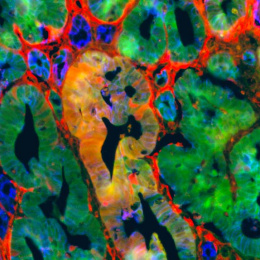
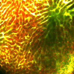
This is an image of a tumor of the bile ducts called cholangiocarcinoma, surrounded by normal liver tissue. The spherical/cuboidal cells (depicted in green or red) are cholangiocytes, the main cells of this tumor type. The interconnected patchwork at the center of the tumor (depicted in red or yellow) is a special nano-probe that we have developed that highlights the presence of proteases, a type of enzyme that is used by cancers to grow, recruit blood vessels, and metastasize. The cells with only the blue stain for nuclei are mostly hepatocytes, the main cells of the liver.
We took this image in order to understand whether our protease-detecting nano-probes were able to detect proteases in cholangiocarcinoma, and which cells in these tumors were responsible for producing proteases.