Spatial Transcriptomic Analysis of Metastatic Cancer 1
Spatial Transcriptomic Analysis of Metastatic Cancer 1
Liangliang Hao, Nour-Saïda Harzallah, and Carmen Martin-Alonso (Bhatia lab); Jonathan Braverman (Yilmaz lab), George Eng (Yilmaz lab), Kathy Cormier (histology core), Jeffrey Kuhn (microscopy core), Vincent Butty (BMC core)
Koch Institute at MIT
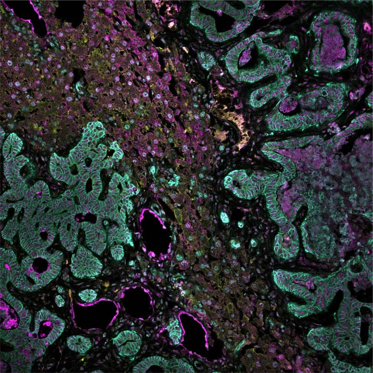
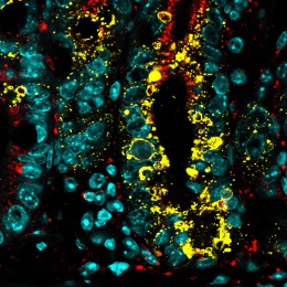
More than 90% of all cancer-related deaths are caused by metastasis, the spread of cancer to a different part of the body than where it started. In this image, we capture disseminated colorectal cancer (CRC) invading the liver, the primary metastatic organ of this tumor type. To invade into their destination organ, cancer cells utilize a class of enzymes, called proteases. We stain the cancer cells (the wave-like granular structures in Cyan, characteristic of intestinal epithelial cells) and the dysregulated proteases (in magenta, red & yellow) in a liver tissue section, to highlight the tumor’s ‘weapons’ of choice during liver invasion. In particular, this image shows the granular tumors invading into the liver space (cells align the top-left/bottom right diagonal). Our overarching research goal is to leverage locally expressed, dysregulated protease activity to develop precision diagnostics and therapeutics for cancer metastasis.