Distribution of A Gene-Delivered Fluorescent Reporter 2
Distribution of A Gene-Delivered Fluorescent Reporter 2
Brent E. Fitzwalter
Dr. Myriam Heiman Laboratory
MIT Department of Brain and Cognitive Sciences, Picower Institute for Learning and Memory
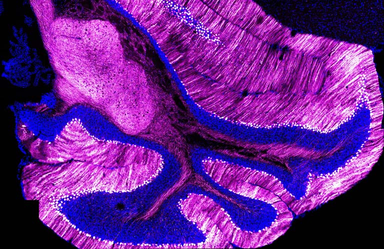
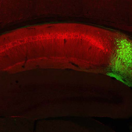
We are testing a gene delivery system to determine if we can insert a fluorescent reporter into specific brain cells called Purkinje neurons. This image shows that the Purkinje neurons—lined up side-by-side, each with their own ‘tree-like’ structure—have received the reporter and display a strong fluorescent signal. Although Purkinje neurons have been studied for nearly 200 years, they still fascinate scientists today due their strikingly beautiful, elaborate branches. Importantly, these cells die in specific brain diseases, leading to movement disorders like Huntington’s disease and spinocerebellar ataxias. Additionally, the cerebellum—the brain structure shown in this image—is the most common site of presentation of central nervous system tumors in children. Having the ability to specifically deliver protective genes to Purkinje neurons may be a viable therapeutic strategy for treating these devastating neurological diseases.
This image was taken to learn how well a gene delivery system is able to distribute a Magenta fluorescent reporter to a specific brain cell type called the Purkinje neuron. It is the first image of many and provides a proof-of-principle that we are able to deliver genetic material—in this case a fluorescent reporter—in a cell type-specific manner. This system will be used in future experiments to lower the levels of a toxic gene that is known to cause cerebellar ataxia. Stay tuned.