A Tri-culture Organoid System Develops in a Synthetic Hydrogel 2
A Tri-culture Organoid System Develops in a Synthetic Hydrogel 2
Alex Wang, Allysa Allen
MIT Department of Biological Engineering, Koch Institute at MIT
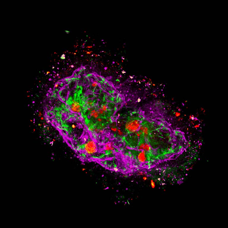
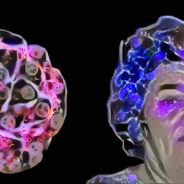
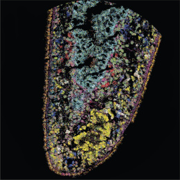
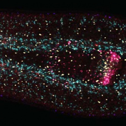
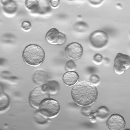
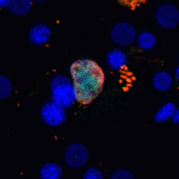
A major goal of liver tissue engineering is to build healthy, vascularized liver tissue in vitro, which will allow us to create larger and more accurate biological models. These series of images depict human liver hepatocyte spheroids (red, albumin), encapsulated in a PEG hydrogel with induced pluripotent stem cell (iPS) derived endothelial cells (green, CD31) and bone-marrow derived mesenchymal stem cells (MSCs) (magenta, vimentin). The combination of iPS endothelial cells and MSCs forms a stable, multi-week sustaining vascular network that interacts with the hepatic spheroids. Over several weeks, this complex system of different cell types proceeds to contract, deform, and degrade their hydrogel microenvironment to form an intriguing organization.
The outer ring of speckled colors most prominent in [this image] is an ephemeral remnant of degraded hydrogel and dead, rounded cells that still show bright coloring. These series of images were taken to visualize the distribution of the different cell types, namely the formation of the vascular network, its evolution over time, and its interaction with the spheroids.
