Visuailizing Vasculature 3
Visuailizing Vasculature 3
Genevieve Abbruzzese, Jeffrey Kuhn, Thao Nguyen, Richard Hynes
MIT Department of Biology, Koch Institute at MIT
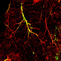
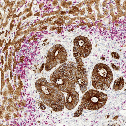
This image is of a mouse retina, highlighting the network of blood vessels (shown in green) that supply the retina with oxygen and nutrients. These vessels grow out radially from the center to the outer edge of the retina. This process of angiogenesis, or growth of the vasculature, is tightly controlled in the retina and is critical for proper eyesight. Defects can lead to blindness in premature infants when the vasculature has not developed properly, or loss of vision in adults when the vessel network is altered by conditions such as diabetes.
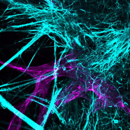
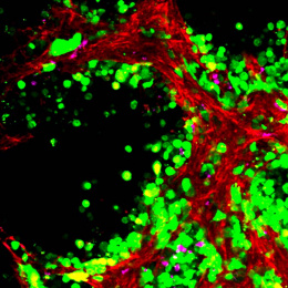
The organized development of vasculature is broadly important in the retina and other tissues, but is also an important component of cancer as the tumor creates its own network of vessels to fuel its growth and metastasis. During both developmental and pathological angiogenesis, the endothelial cells that form the walls of blood vessels make contacts with and receive signals from the surrounding extracellular matrix (ECM) in their microenvironment. This promotes and controls their growth and migration. We took this image of a retina to visualize the vasculature and underlying ECM. We want to better understand how specific ECM proteins that organize into a fibrous meshwork beneath the developing vessels guide the growing vessel network and promote proper angiogenesis.

