Trafficking Nanoparticles to the Lymph Nodes 3
Trafficking Nanoparticles to the Lymph Nodes 3
Ben Read, Jason Chang
MIT Department of Biological Engineering, MIT Department of Materials Science and Engineering, Koch Institute at MIT
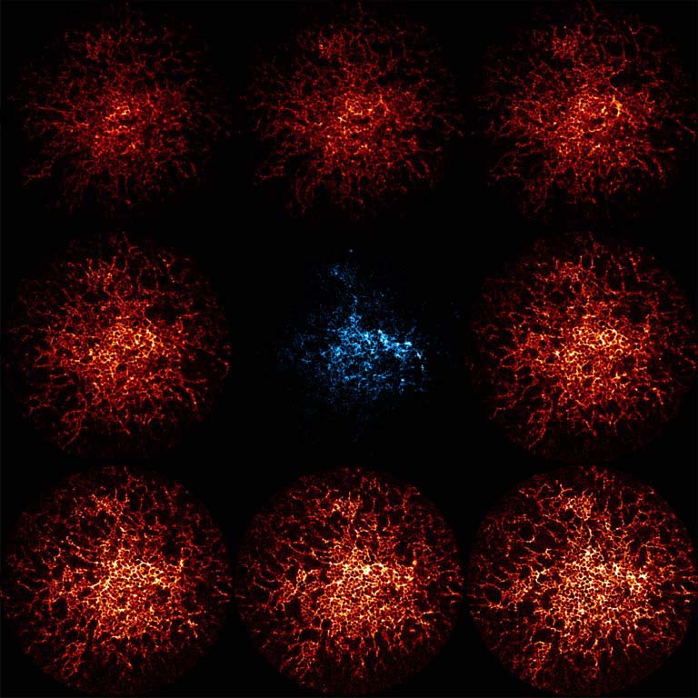
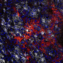
This image shows part of a germinal center, the training ground of the immune system. The wider spread that can be seen are cells (follicular dendritic cells) that display foreign materials so other elements of the immune system can test if they can recognize the specific foreign substance. The color concentrated more in the center is a small protein nanoparticle (ferritin) that has been coated in mannose, a simple sugar, which causes the particle to traffic rapidly to follicular dendritic cells as part of a newly elucidated mechanism.
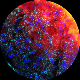
This image was taken to determine if our modification to the ferritin nanoparticle would cause it to be displayed by follicular dendritic cells. The outer ring of images represents the follicular dendritic cells “moving” up through the Z dimension. The inner image is the ferritin signal averaged throughout the Z dimension.
