HIV Vaccine Visualized Inside a Lymph Node 2
HIV Vaccine Visualized Inside a Lymph Node 2
Jacob T. Martin
MIT Department of Biological Engineering, MIT Department of Materials Science and Engineering, Koch Institute at MIT
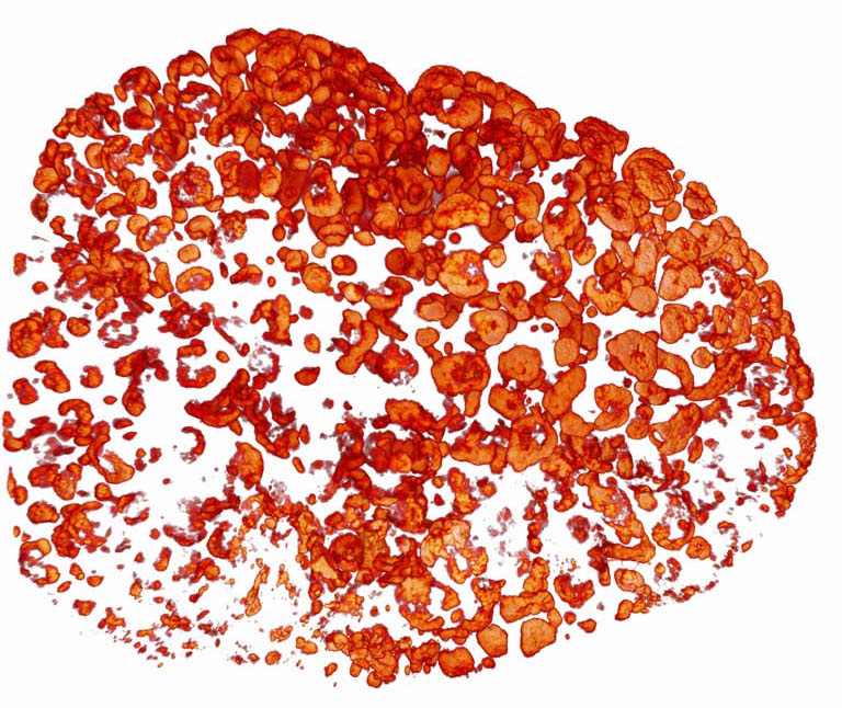
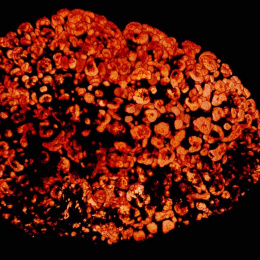
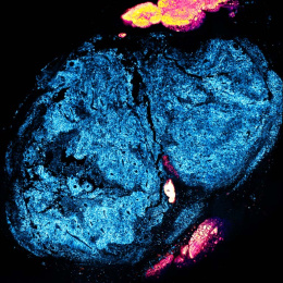
This image shows HIV vaccine visualized in 3D inside a lymph node. Lymph nodes are the organs in which the immune system learns to defend the body against pathogens. We are trying different ways to get as much of our HIV vaccine as possible to the lymph nodes so that the immune system can develop a stronger response. Visualizing this transport helps us determine which vaccination strategies work best.
By taking advantage of recently-developed methods for clearing and visualizing molecules in whole organs (as opposed to those which have been sliced up for histology), we are able to see the three-dimensional migration of our vaccine components. We are finding new patterns of antigen deposition in lymph nodes that may not have ever been appreciated by histological sectioning and imaging alone.
