Examining Organoids, Image 3
Examining Organoids, Image 3
Marit van Gorsel, Daniel Schmidt, Matthew Vander Heiden
MIT Department of Biology, Koch Institute at MIT
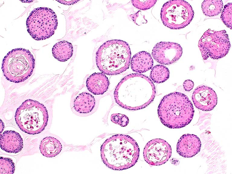
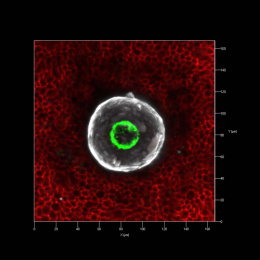
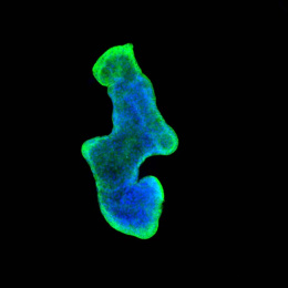
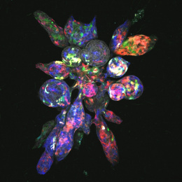
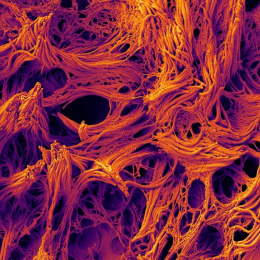
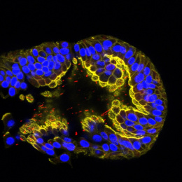
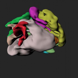
These pictures all show normal mouse prostate organoids, 3D spheres of cells that grow in a culture dish. We took these images to keep track and to study the phenotype of these organoids. The H&E stained organoids (first picture), gives a beautiful insight in their structure. The organoid culture system allows us to culture normal prostate cells, which is very hard in 2D tissue culture. Since this is such a new culture system, it was very important to get to know the behavior and the structure of these organoids.
