Getting Into C. elegans' Head 2
Getting Into C. elegans' Head 2
Rita Droste, Nikhil Bhatla
MIT Department of Biology, McGovern Institute for Brain Research, Koch Institute at MIT
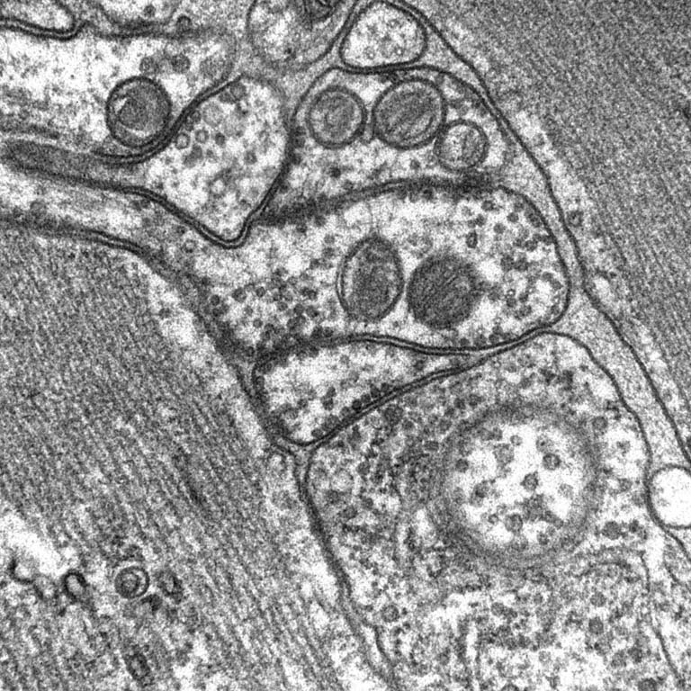
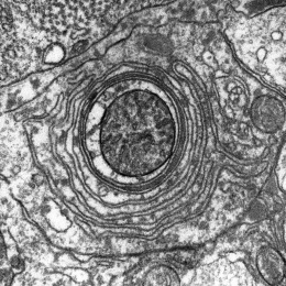
This is an image of a slice of the roundworm C. elegans' head, taken using an electron microscope. In the center of the image, you can see several circles sitting in a row at the cellular membrane. These are synaptic vesicles, and they contain neurotransmitters which serve as the medium of communication between neurons. These vesicles are primed and ready to be released instantly when the neuron becomes activated, and they control the feeding behavior of the worm. These neurons are sandwiched between two muscles, whose fibers can be seen streaming by on either side.
This image was taken in the context of a serial section reconstruction of the worm's feeding organ, the pharynx. We were studying the synaptic connectivity between neurons and muscle visible only by electron microscopy.
