Sphere, There, and Everywhere: Interrogating Tiny Tumors
Sphere, There, and Everywhere: Interrogating Tiny Tumors
Collections: Image Award Winners
2016 Award Winner
Alexandre Albanese, Jeffrey Wyckoff, Sangeeta Bhatia
Koch Institute at MIT, Institute of Medical Engineering and Science
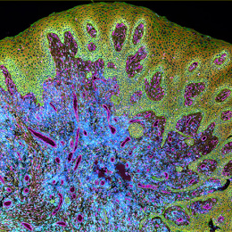
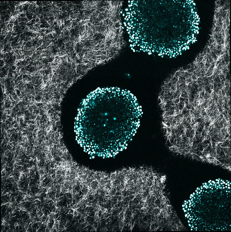
Cancer cell experiments fall flat at the bottom of a Petri dish. This image shows three spherical clusters of cancer cells (blue/white dots) implanted in a three-dimensional matrix of protein fibers (white strands).
These tiny tumors allow researchers to understand how small metastases interact with their surrounding environment, and to evaluate the delivery of drug-loaded nanoparticles. The use of spheroids rather than isolated cells offers a more well-rounded picture of the challenges researchers must overcome to detect and treat cancer.
Video
Alex Albanese shares the story behind his award-winning image.