Neurons Divide and Mature, Image #1
Neurons Divide and Mature, Image #1
Submitted by Kristin Knouse in the Amon Laboratory at the Koch Institute
MIT Department of Biology, Koch Institute at MIT
Kristin Knouse
Amon Lab, Koch Institute
Spinning Disc Confocal Microscopy
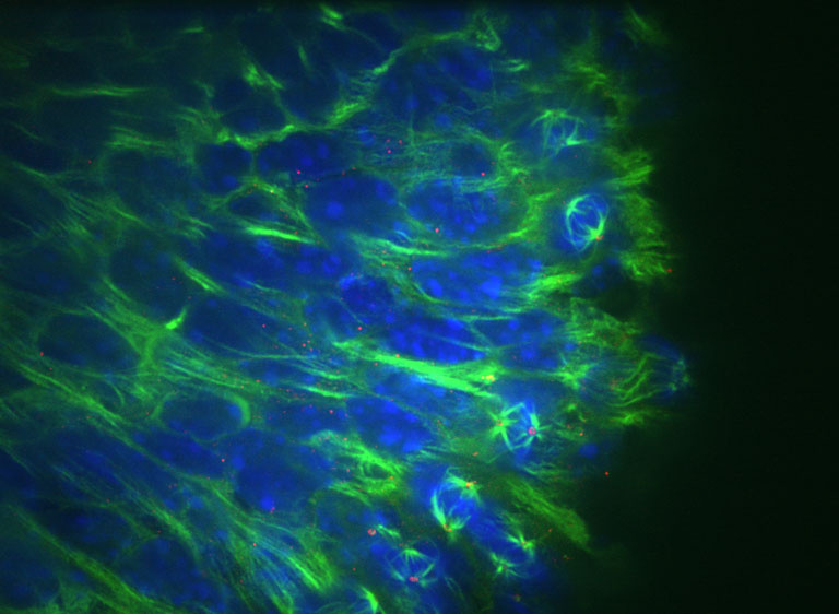
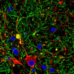
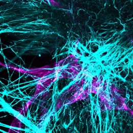
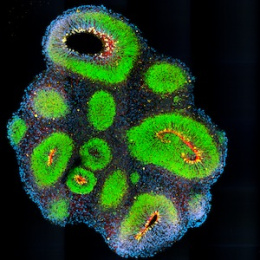
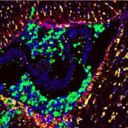
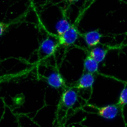
"These images are taken from brain tissue of embryonic mice. The cells at the base represent neural stem cells, and at this period they are actively dividing to give rise to mature neurons that will remain with the animal through adulthood. We are interested in whether abnormal cell division in neural stem cells leads to aberrant chromosome number in neurons. These images allow us to analyze aspects of cell division in the neural stem cells and determine whether or not cell division is occurring normally.
Our laboratory studies various aspects of chromosome segregation and chromosome imbalance. We suspect that the brain may have increasing levels of chromosome missegregation, and our analysis of cell division in these images will help us understand the prevalence and mechanism of this defect."
