Mechanisms of Retinal Degeneration, Version #2
Mechanisms of Retinal Degeneration, Version #2
Submitted by Jennifer Calvo, Lisianne Meira, Catherine Moroski and Leona Samson of the Samson Laboratory in the Center for Environmental Health Sciences and the Department of Biological Engineering
MIT Department of Biological Engineering, Koch Institute at MIT
Jennifer Calvo, Lisianne Meira, Catherine Moroski, Leona Samson
Samson Laboratory, Center for Environmental Health Sciences and Department of Biological Engineering
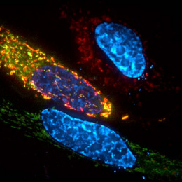
Epi-Fluorescence Micrograph
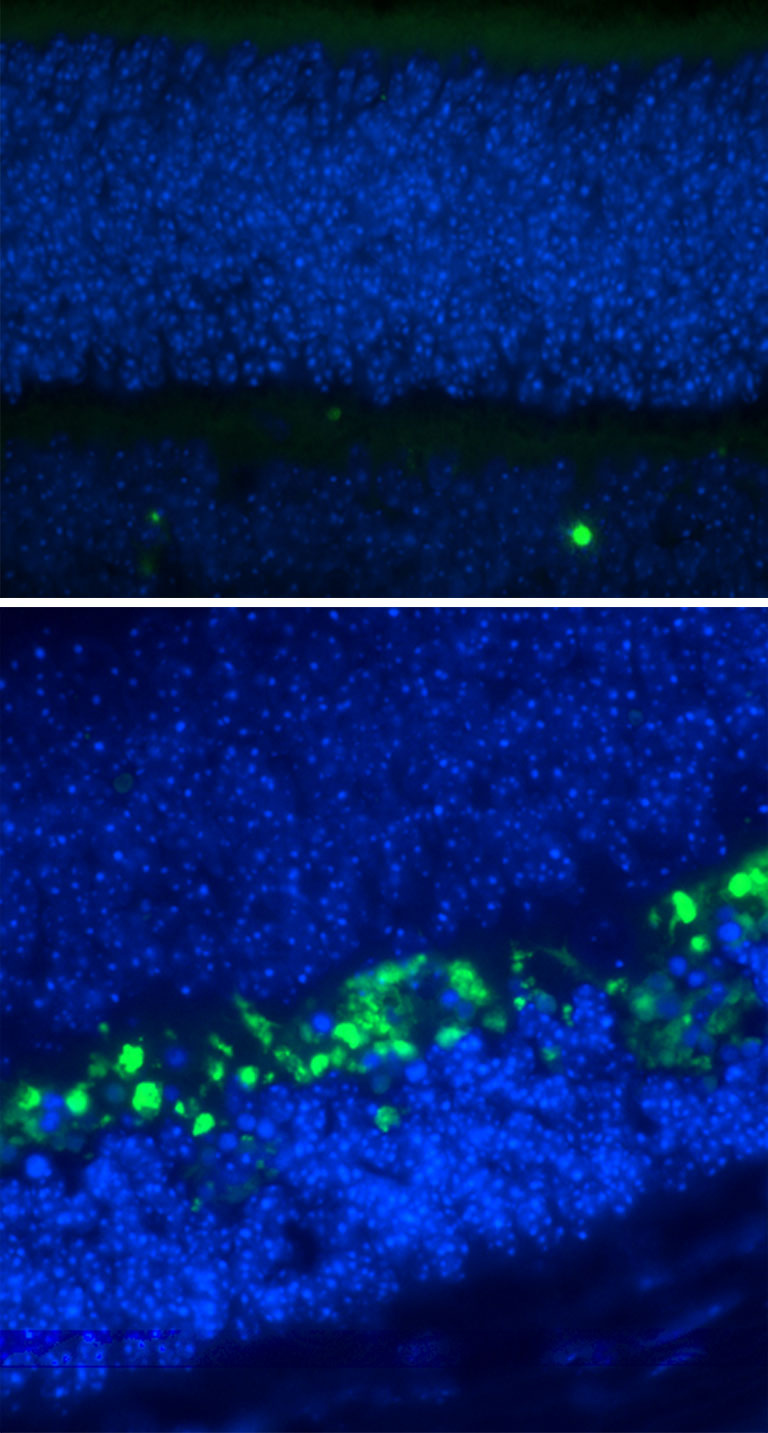
"Vision loss affects more than 3 million Americans and many more people worldwide. Although predisposing genes have been identified their link to known environmental factors is unclear. Shown here is an immunofluorescence picture of retinas isolated 24 hours following treatment with the alkylating agent MMS. TUNEL (green) staining indicates cells undergoing apoptosis (cell death), and DAPI staining illustrates the nuclei of cells. The retina lacking alkyladenine DNA glycosylase (AAG) exhibits no apoptosis (top image). A wild-type retina displays significant apoptosis, specifically in the outer nuclear layer of the retina (bottom image). The image demonstrates that AAG may contribute to alkylating agent-induced retinal degeneration."
